
Ureteroscopic lithotripsy (URS)
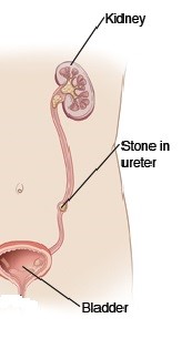

Ureter stone and its appearance on xray
This is the endoscopic treatment of ureter stones using a mini-scope and Holmium laser to break the stone.
How it is done
The surgery can be done as a day case under general anaesthesia and xray guidance, A rigid or flexible ureteroscope is passed up the ureter via the urethra to reach the stone. The Holmium laser is passed through the scope to break the stone into smaller pieces. These tiny stone pieces will pass out on their own. A wire basket can also be used to trap the stone or extract the broken stones. This surgery takes 30 mins on average.

Rigid vs flexible ureteroscope
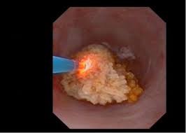
Holmium laser to break the stone
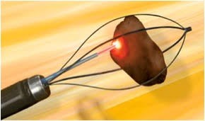
Dormia basket to trap / extract the stone
Watch video on how stone laser is done in the ureter with the rigid ureteroscope
Watch video on stone laser is done in the kidney with the flexible ureteroscope
Occasionally, a double-J (DJ) stent may need to be inserted after the procedure should there is any injury to the ureter wall or if there is prior gross hydronephrosis of the kidney caused by the impacted stone. The DJ stent can be removed 2 to 3 weeks later under local anaesthesia using a flexible cystoscope. The success rate for stones lodged in the lower ureter is near 100%. For stones lodged at the mid- to upper ureter, there is a chance they may float up into the kidney. If the flexible ureteroscope is used, this is not an issue as the scope can go into the kidney calyces to find the stone should it float into the kidney. However, if the rigid ureteroscope is only available, then the floated-up stone cannot be treated and a DJ stent is inserted for subsequent ESWL.
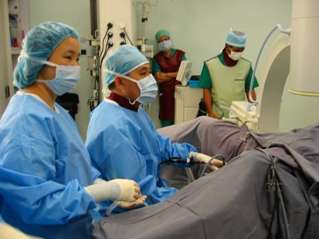
Dr Chin doing URS surgery
The advantages of URS over ESWL is that even hard stones can be broken with the laser and the ureter opening can be dilated to facilitate subsequent stone passage. However, URS is a more expensive and more invasive with the real risk of injuring or even avulsing the ureter.
Complications include:
- bloody urine. This should clear up in a few days
- This occurs post-operatively and is due to bacteria released from the stone as it breaks. This can be minimised by giving antibiotics prior to surgery
- perforation of the ureter. If this happens, urine leak, fever and pain results. A DJ stent will need to be inserted to prevent further urine leak and avoid a late stricture
- stone migration. Because pressurised water is used to access the ureter to visualise the stone, the pressure may accidentally push the stone up beyond the reach of the ureteroscope
Desired outcomes:
- no ureter injury
- complete stone clearance
- no need to insert a DJ stent ( in 95% of cases )